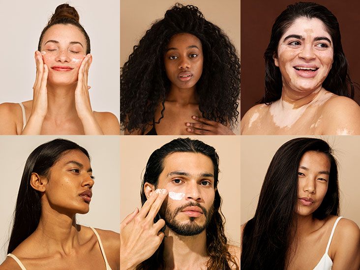
In medicine, we love a neat little box. Check the box, click the dropdown, move on with our lives.
And when it comes to skin, we’ve been trying to tidy up something gloriously untidy for decades.
“What’s your skin type?” sounds simpleuntil you realize we’re using one tool for sunburn risk, another for skin tone,
and a third for how redness shows up… and then we act surprised when patients don’t fit the script.
This is a doctor’s plea (with mild caffeine-fueled side-eye): let’s honor diversity in skin typing.
Not as a buzzword, but as a clinical necessity. Because when our labels are sloppy,
our care can get sloppy tooespecially for patients with darker skin tones who’ve been underserved by
“classic presentations” and underrepresented images for far too long.
Skin Typing Isn’t Just Dermatology Nerd StuffIt’s Patient Safety
Skin typing shows up everywhere: dermatology, primary care, aesthetics, emergency medicine,
device approvals, clinical trials, even the training slides we show students at 7 a.m. (cruel, but traditional).
We use it to estimate sunburn risk, counsel on skin cancer prevention, choose laser settings,
anticipate hyperpigmentation, and dose phototherapy.
But here’s the twist: the most widely used frameworkthe Fitzpatrick Skin Type (often called “phototype”)
was created to predict how skin responds to ultraviolet light, not to serve as a universal stand-in for
skin color, race, or ethnicity. In practice, it’s frequently stretched beyond its original purpose,
and that stretch can snap into real-world inequities.
The Fitzpatrick Scale: Useful Tool, Misused Shortcut
The Fitzpatrick system (I–VI) is best thought of as a UV-response scale:
how likely you are to burn or tan. It’s clinically helpful for discussing sun sensitivity and some
treatment planning. The trouble starts when we treat it like a “skin color ruler.”
Two people can share a similar visible skin tone but have different burn/tan histories.
And clinicians may “estimate” a patient’s type based on appearanceoften inaccuratelyespecially
in diverse populations.
Where the shortcut hurts
-
Missed or delayed diagnoses: In darker skin, inflammation and redness may not look
“pink” or “bright red.” It can appear violaceous, gray-brown, or subtleso conditions like dermatitis,
infections, or even drug reactions may be underestimated if we’re trained on a narrow visual library. -
Skin cancer myths: People with darker skin can and do get skin cancer.
Some types may be diagnosed later, with worse outcomes, partly because of under-recognition and
the persistent cultural myth that melanin makes you “immune.” -
Procedural risks: Lasers, peels, and other procedures can carry higher risks of
post-inflammatory hyperpigmentation in some patients. Using an imprecise label can mean settings that
are too aggressiveor a patient being discouraged from helpful treatments out of exaggerated fear.
When we compress a person into a single number without context, we lose clinically meaningful nuance:
undertones, baseline pigmentation, tanning practices, scarring tendencies, history of pigment changes,
and how erythema actually shows up on their skin.
Representation Gaps: When “Classic” Means “Only on Light Skin”
Medical education has a long-running problem: too many “classic” textbook photos and teaching cases
skew toward lighter skin. If trainees rarely see psoriasis, Lyme rashes, eczema, or cellulitis on
darker skin, they’re set up to miss it in real life. That’s not just a diversity issueit’s a diagnostic
accuracy issue.
More inclusive image libraries and teaching materials aren’t cosmetic upgrades; they are patient-care
upgrades. When clinicians learn to recognize disease across a wide range of skin tones, fewer people
get told, “Huh, that’s weird,” when what they really need is timely treatment.
Beyond Fitzpatrick: Skin Tone Scales, Color Tools, and Better Documentation
If Fitzpatrick is about UV response, what about documenting skin tone itself?
Increasingly, clinicians and researchers are exploring complementary approaches:
scales designed to represent a broader spectrum of skin tones (rather than sunburn response),
and methods that are more objective than “eyeballing it.”
The rise of broader tone scales
One important development is the Monk Skin Tone (MST) scale, which offers more gradations of skin tone
than older tools and aims to be more representative. It’s been discussed as a way to improve how we
label skin tone in research, imaging datasets, and clinical communicationespecially in contexts where
“tone” (not “burn/tan behavior”) is the variable that matters.
No scale is perfect. Printing, lighting, and display differences can affect tone matching.
But the broader point stands: if we’re documenting skin tone, we should use a tool built for skin tone
and we should be transparent about its limitations.
Practical clinic moves that work today
-
Ask the patient, don’t guess: For Fitzpatrick phototype, ask about burn/tan history.
For tone concerns (hyperpigmentation risk, device accuracy concerns), ask about prior pigment changes,
scarring, and what “redness” looks like for them. -
Document both “phototype” and “tone” when relevant: A patient may have a phototype
that reflects UV response and a skin tone description that helps with visual assessment and procedural planning. -
Use consistent lighting: If your clinic takes photos, standardize lighting and background.
Variable lighting is how “before-and-after” turns into “before-and-who-is-that.” -
Describe what you see in plain language: Instead of “erythematous rash,” consider:
“ill-defined patches with warmth and swelling; color appears deep red-brown/violaceous on this patient’s skin.”
Device Bias Isn’t Abstract: Pulse Oximetry and the Cost of Oversimplified Skin Categories
If you want a crystal-clear example of why skin pigmentation matters beyond dermatology, look at pulse oximetry.
Research and policy discussions have highlighted that oximeter accuracy can vary with skin pigmentationan issue
that gained major attention during the COVID-19 era. The FDA has issued updated draft recommendations aimed at
improving pulse oximeter performance across a range of skin pigmentations and strengthening study diversity expectations.
Here’s the lesson for skin typing: when skin tone is treated as an afterthoughtor measured inconsistently
it can translate into clinical blind spots. Not because anyone wakes up hoping to build inequity into a device,
but because “we tested on whoever showed up” often means “we tested on a narrow slice of humanity.”
AI and Dermatology: If the Dataset Is Pale, the Predictions Will Be Too
AI tools in dermatology are expanding fast: lesion classifiers, rash triage apps, image-based decision support.
But AI learns patterns from data. If datasets underrepresent darker skin tones, the model can become an expert
in diagnosing disease on light skinand a rookie on everyone else.
This is where better skin tone labeling matters: not as a checkbox, but as a foundation for fairness and accuracy.
Reliable labeling helps researchers audit representation, measure performance across groups, and reduce preventable bias.
The goal isn’t to make every patient fit a tidy category; it’s to make our tools work for actual patients.
So What Does “Honoring Diversity in Skin Typing” Look Like?
It looks like refusing to pretend one number captures a person. It looks like teaching students that
“classic” is not synonymous with “light skin.” It looks like documenting skin thoughtfully when it mattersand
not using skin labels as a proxy for race.
Three principles I wish we taught on day one
- Purpose first: Choose the tool that matches the clinical question (UV response vs. tone vs. erythema visibility).
- Patient partnership: Let patients describe their lived experience of sun response, pigment change, and what “redness” looks like.
- Humility over habit: If you’ve always done it a certain way, that’s not proof it’s the best wayjust proof you’ve been busy.
What I Tell Patients (Because They Deserve the Truth, Not the Myth)
I tell patients with darker skin: you still need sun protectionnot because you’re fragile, but because UV contributes
to pigment changes, photoaging, and skin cancer risk in every skin tone. I tell patients with lighter skin:
“burning is not a personality trait; it’s your skin asking for backup.” And I tell everyone: if a spot changes,
bleeds, doesn’t heal, or looks “off,” it deserves attentionno matter your skin tone.
I also tell patients that medicine is catching up to what they’ve always known: their skin doesn’t read the textbook,
and neither should their care.
In the Exam Room: of Lived Lessons on Skin Typing
I learned the limits of skin typing the way many doctors do: one humbling patient at a time.
Early in training, I was taught to look for “erythema” like it was a neon sign. Red means inflamed. Red means urgent.
Simpleuntil I met a patient whose severe dermatitis showed up as a deep, muted purple-brown. The skin was hot,
thickened, and intensely itchy, but it didn’t scream “red” the way my mental flashcards expected.
My first thought wasn’t my proudest: “It doesn’t look that bad.” The patient’s face said,
politely, that I was wrong. The patient’s sleeplessness said it louder.
Another moment: a young adult came in worried about “dark patches” after acne healed. Their previous clinician had
shrugged and called it “just scarring.” But it wasn’t scarring in the fibrotic sense; it was post-inflammatory
hyperpigmentationsomething that can be profoundly distressing, especially when it’s dismissed.
When we talked through their history, the pattern was clear: every flare left a mark, and every mark lingered longer
than their friends’ did. That conversation changed how I document skin. Instead of slapping on a label and moving on,
I started writing what mattered: what happens after inflammation, what triggers it, what products make it worse,
what helps, and what the patient fears. Skin typing became less about numbers and more about outcomes.
Then there’s the cosmetic consult that taught me the ethical side of this topic. A patient with richly pigmented skin
wanted a laser treatment. Another office had told them, “You’re not a candidate,” full stopno explanation, no nuance.
When I reviewed their history, I saw a patient who understood risks, had stable skin, and had realistic goals.
The right move wasn’t a blanket “yes” or “no.” It was a risk-balanced plan: conservative settings, test spots, sun protection,
close follow-up, and an honest discussion of possible pigment change. They did well. The bigger win wasn’t the outcome
(though they were happy); it was the message: you deserve individualized care, not a reflexive rejection because a scale
wasn’t designed to hold your complexity.
I’ve also watched colleagues get betterfastonce they realize the “classic” images they memorized were incomplete.
When we started sharing better photo references and discussing how conditions present across tones, diagnostic confidence
improved. The room felt different. Less guesswork. Less awkward “Hmm.” More accurate medicine.
So yes, I still use Fitzpatrick phototypes when they’re relevant. But now I treat them like one tool in a larger kit.
Honoring diversity in skin typing, to me, means listening carefully, documenting thoughtfully, and refusing to let a
simplified system shrink a patient’s reality. The skin is our largest organ. Our understanding of it should be big too.
Conclusion: Better Skin Typing Is Better Care
We don’t need to throw out old toolswe need to use them correctly and stop pretending they do jobs they were never built to do.
When we honor diversity in skin typing, we reduce missed diagnoses, improve procedural safety, strengthen research quality,
and build trust with patients who are tired of being told their skin is an “exception.”
Skin typing should never be a shortcut around curiosity. It should be a prompt to ask better questionsand to practice
medicine that fits the person in front of us.