Note: This article is for educational purposes only and should not replace medical advice. Anyone with bone pain, height loss, fractures, long-term steroid use, or concerns about bone density should speak with a qualified healthcare professional.
Osteoporosis is often described as a “silent disease,” which sounds dramatic enough to be the title of a mystery novel. But the nickname fits: bones can weaken for years without causing obvious symptoms, and the first clue may be a fracture from a fall that did not seem serious enough to break anything. Secondary osteoporosis adds another twist to the story. It means bone loss is not only happening because of age, menopause, or natural bone changes, but because another condition, medication, hormone problem, or lifestyle factor is pushing the skeleton in the wrong direction.
Understanding the difference between primary and secondary osteoporosis matters because the treatment plan may change completely. If the real problem is untreated hyperthyroidism, celiac disease, chronic kidney disease, low testosterone, long-term glucocorticoid use, or severe vitamin D deficiency, simply telling someone to “drink more milk” is like trying to fix a leaking roof with a decorative umbrella. Helpful? Maybe for vibes. Not enough for the actual problem.
This guide explains what secondary osteoporosis is, how it differs from primary osteoporosis, what causes it, how doctors diagnose it, and why a careful evaluation can protect more than bonesit can protect independence, mobility, and quality of life.
What is secondary osteoporosis?
Secondary osteoporosis is osteoporosis caused or worsened by another identifiable factor. That factor may be a medical condition, a medication, a hormonal disorder, a nutritional problem, or a chronic disease that interferes with how the body builds, maintains, or repairs bone.
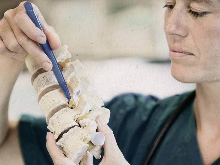
In healthy bone, the body constantly removes old bone tissue and replaces it with new tissue. This remodeling process is normal. Think of it as a construction crew that never clocks out. Osteoporosis develops when bone breakdown outpaces bone formation, leaving bones less dense, more fragile, and more likely to fracture. In secondary osteoporosis, something external to normal aging disrupts that balance.
For example, long-term use of glucocorticoids such as prednisone can reduce bone formation and increase fracture risk. Hyperparathyroidism can pull calcium from bones. Celiac disease may reduce nutrient absorption, including calcium and vitamin D. Rheumatoid arthritis can increase inflammation and reduce mobility. Chronic kidney disease can affect mineral balance. Some cancer treatments can lower sex hormones that help preserve bone strength. The result is the same general destinationweaker bonesbut the route is different.
Primary vs. secondary osteoporosis: the key difference
The simplest way to compare primary and secondary osteoporosis is this: primary osteoporosis is usually related to aging and natural hormonal changes, while secondary osteoporosis has a specific underlying trigger.
Primary osteoporosis
Primary osteoporosis is the most common form. It often develops after menopause because estrogen levels drop, and estrogen plays a major role in maintaining bone density. It also occurs with aging in both women and men as bone rebuilding slows down. Over time, bones may lose mineral density and structural strength.
Common risk factors for primary osteoporosis include older age, family history of osteoporosis, low body weight, previous fragility fracture, smoking, heavy alcohol use, low calcium intake, low vitamin D, and limited weight-bearing activity. Primary osteoporosis is not “someone’s fault.” Bones are living tissue, and they age like the rest of the bodyalthough, admittedly, bones are less likely to complain on Monday mornings.
Secondary osteoporosis
Secondary osteoporosis can occur at any age. It may appear in postmenopausal women, older men, younger adults, and sometimes even people who seem too young or healthy to have low bone density. That is why doctors often look more closely when osteoporosis appears unexpectedly, such as in a premenopausal woman, a man under 50, or a person with multiple fractures without major trauma.
Secondary osteoporosis does not always replace primary osteoporosis; it can overlap with it. A 68-year-old woman may have age-related bone loss and also be taking long-term steroids for an autoimmune condition. A 72-year-old man may have age-related bone loss plus low testosterone or vitamin D deficiency. In real life, bones do not politely choose one category for the convenience of medical textbooks.
Common causes of secondary osteoporosis
Secondary osteoporosis has many possible causes. Some are common, some are sneaky, and some are discovered only after a fracture or abnormal bone density test leads to a deeper evaluation.
Medication-related causes
Certain medications can weaken bones when used for a long time or at higher doses. Glucocorticoids are one of the best-known examples. These drugs can be essential for conditions such as asthma, lupus, inflammatory bowel disease, or rheumatoid arthritis, but they can also reduce bone formation.
Other medications that may affect bone health include some anti-seizure medicines, aromatase inhibitors used in breast cancer treatment, androgen deprivation therapy used in prostate cancer treatment, some drugs that suppress hormones, and long-term use of certain acid-reducing medications in specific risk settings. Not everyone taking these medicines will develop osteoporosis, but they are worth discussing with a clinician.
Hormonal and endocrine disorders
Hormones are bone’s behind-the-scenes managers. When they go off script, bones notice. Hyperthyroidism, hyperparathyroidism, Cushing syndrome, diabetes, low estrogen, low testosterone, and pituitary disorders can all contribute to bone loss.
Low sex hormone levels are especially important. Estrogen and testosterone help maintain bone remodeling balance. When these hormones fall early or sharplybecause of menopause, certain cancer treatments, eating disorders, pituitary disease, or testicular conditionsbone density may decline faster than expected.
Digestive and absorption disorders
Bones need raw materials, especially calcium, vitamin D, protein, magnesium, and phosphorus. If the digestive system cannot absorb nutrients properly, bone strength can suffer. Celiac disease, inflammatory bowel disease, bariatric surgery, chronic liver disease, and pancreatic disorders may all affect nutrient absorption.
This is one reason secondary osteoporosis can be missed. A person may eat a reasonably healthy diet but still not absorb enough nutrients. The skeleton is not impressed by good intentions; it wants delivery confirmation.
Inflammatory and autoimmune diseases
Chronic inflammation can accelerate bone loss. Rheumatoid arthritis, lupus, ankylosing spondylitis, inflammatory bowel disease, and other inflammatory conditions may increase osteoporosis risk. These conditions may also reduce physical activity, increase fall risk, or require medications that affect bone density.
Kidney, liver, and blood disorders
Chronic kidney disease can disrupt calcium, phosphorus, vitamin D, and parathyroid hormone balance. Chronic liver disease can interfere with vitamin D metabolism and nutrition. Blood disorders such as multiple myeloma can directly weaken bones and increase fracture risk.
When osteoporosis is severe, unusual, or accompanied by anemia, unexplained weight loss, high calcium levels, kidney problems, or persistent pain, doctors may investigate more serious underlying causes.
Symptoms: why secondary osteoporosis can be hard to spot
Osteoporosis often has no symptoms until a fracture occurs. That is true for both primary and secondary osteoporosis. A person can feel completely normal while bone density is quietly dropping. No flashing dashboard light. No dramatic theme music. Just bones doing their best under difficult circumstances.
Possible signs that deserve medical attention include:
- A fracture from a minor fall or low-impact injury
- Loss of height over time
- Stooped posture or new spinal curvature
- Sudden back pain, especially if related to a vertebral compression fracture
- Low bone density on a DXA scan
- Osteoporosis at a younger-than-expected age
Secondary osteoporosis may also come with symptoms of the underlying cause. For example, hyperthyroidism may cause weight loss, heat intolerance, anxiety, or a fast heartbeat. Celiac disease may cause digestive symptoms, fatigue, anemia, or no obvious symptoms at all. Low testosterone may cause low energy, reduced muscle mass, or changes in sexual function. The clue is not always in the bones alone.
How doctors diagnose osteoporosis
The main test used to diagnose osteoporosis is a bone density test, most often a DXA scan. DXA stands for dual-energy X-ray absorptiometry. It is quick, noninvasive, and commonly measures bone density at the hip and spine. These areas matter because hip and vertebral fractures can have major effects on mobility and long-term health.
Understanding T-scores
DXA results often include a T-score. A T-score compares a person’s bone density with that of a healthy young adult. In general:
- A T-score of -1.0 or above is considered normal bone density.
- A T-score between -1.0 and -2.5 is called low bone mass, also known as osteopenia.
- A T-score of -2.5 or lower is consistent with osteoporosis.
A diagnosis may also be made if a person has a fragility fracture, particularly of the hip or spine, even if the T-score does not tell the whole story. Bone density is important, but fracture history matters too.
Understanding Z-scores
A Z-score compares bone density with what is expected for someone of the same age, sex, and body size. Z-scores are especially useful in younger adults, premenopausal women, and men under 50. A very low Z-score may raise suspicion for secondary osteoporosis, because it suggests bone density is lower than expected for that person’s age group.
How secondary osteoporosis is diagnosed
Diagnosing secondary osteoporosis is not just about proving that bones are weak. It is about asking, “Why are they weak?” That question is where the detective work begins.
Medical history
A clinician will usually review fracture history, family history, diet, exercise, smoking, alcohol intake, menstrual history, hormone-related symptoms, digestive symptoms, kidney disease, cancer history, and medication use. The medication list is especially important. Patients should include prescription drugs, over-the-counter medicines, supplements, injections, and past long-term treatments.
Physical examination
A physical exam may look for height loss, spinal curvature, balance issues, muscle weakness, thyroid enlargement, signs of hormonal disorders, or clues of chronic disease. Sometimes the exam is normal, which does not rule out secondary osteoporosis.
Laboratory testing
Doctors may order blood and urine tests to look for underlying causes. Common tests may include calcium, phosphorus, kidney function, liver function, complete blood count, thyroid-stimulating hormone, 25-hydroxyvitamin D, parathyroid hormone, and markers of inflammation or malabsorption. In selected cases, testing may include testosterone, celiac disease antibodies, serum protein electrophoresis, urine calcium, or other hormone studies.
The exact test list depends on the patient. A young adult with low bone density, a person with multiple fractures, and an older adult taking steroids may need different evaluations. Good diagnosis is not a fishing expedition; it is targeted detective work with a lab coat.
Imaging for fractures
Some people have vertebral compression fractures without realizing it. If there is height loss, back pain, or a very low T-score, clinicians may recommend spine imaging or vertebral fracture assessment. Finding a silent vertebral fracture can change the risk category and treatment plan.
Who should be evaluated for secondary causes?
Many guidelines recommend considering secondary causes in people with osteoporosis, especially when the pattern is unusual or severe. Evaluation is particularly important for:
- Premenopausal women with low bone density or fragility fractures
- Men with osteoporosis, especially younger men
- Anyone with fractures from minimal trauma
- People with very low bone density
- Patients losing bone despite treatment
- People taking long-term glucocorticoids or other bone-affecting medications
- Patients with symptoms suggesting thyroid, parathyroid, digestive, kidney, liver, inflammatory, or blood disorders
Screening recommendations for osteoporosis often focus on age and sex, such as women age 65 and older and postmenopausal women under 65 with increased fracture risk. But secondary osteoporosis is different from routine screening. It asks whether another medical issue is speeding up bone loss or making fractures more likely.
Why diagnosis matters: treatment depends on the cause
Finding secondary osteoporosis can change treatment in several ways. First, the underlying condition may need direct treatment. For example, correcting vitamin D deficiency, treating celiac disease, adjusting thyroid medication, managing parathyroid disease, or reducing steroid exposure when medically possible may help protect bone.
Second, fracture risk may be higher than the bone density number suggests. A person taking long-term glucocorticoids, for example, may have increased fracture risk even before the T-score reaches the osteoporosis range. Third, some bone conditions can mimic osteoporosis but require different care. That is why a careful diagnosis matters before jumping into treatment.
Treatment may include nutrition changes, vitamin D and calcium optimization, weight-bearing and resistance exercise, fall prevention, medication review, and prescription osteoporosis therapy when appropriate. Common medication classes include bisphosphonates, denosumab, anabolic agents, and others, but the right choice depends on fracture risk, age, kidney function, medical history, and the underlying cause.
Practical examples of secondary osteoporosis
Example 1: The long-term prednisone patient
A 55-year-old woman takes prednisone for an autoimmune condition. She has never broken a bone, but her DXA scan shows low bone density. Her doctor evaluates calcium, vitamin D, kidney function, and fracture risk, then discusses steroid-sparing options with her specialist. Her plan may include bone-protective medication, strength training, fall prevention, and careful monitoring.
Example 2: The younger man with a fragility fracture
A 42-year-old man breaks a wrist after a minor fall. Because osteoporosis is less expected at his age, his clinician orders lab testing. Results show low testosterone and vitamin D deficiency. Treating the underlying issues becomes part of the bone health plan.
Example 3: The “healthy eater” with celiac disease
A 60-year-old woman eats well but has low bone density and iron deficiency. Testing suggests celiac disease, which has reduced nutrient absorption. A gluten-free treatment plan supervised by her healthcare team, along with nutritional support, helps address the cause rather than only the consequence.
Living with secondary osteoporosis: experience-based insights
People dealing with secondary osteoporosis often describe the experience as confusing at first. The diagnosis may arrive after a fracture, a routine DXA scan, or an investigation for another condition. Many patients say their first reaction is surprise: “But I walk every day,” “I eat yogurt,” or “I thought osteoporosis was only something older women get.” That surprise is understandable. Secondary osteoporosis does not always match the stereotype.
One common experience is frustration with uncertainty. A person may receive a low bone density result and immediately wonder what went wrong. The answer is not always obvious. It may take a careful review of medications, several blood tests, and sometimes referrals to endocrinology, rheumatology, gastroenterology, nephrology, or oncology. This can feel like a medical scavenger hunt, except the prize is paperwork and another appointment. Still, the process is valuable because finding a treatable cause can make the care plan more effective.
Another experience is learning that bone health is connected to the whole body. Patients often start with a simple idea“My bones are thin”and then discover the skeleton is influenced by hormones, kidneys, digestion, muscles, medications, inflammation, nutrition, and balance. This bigger picture can be empowering. Instead of seeing osteoporosis as a random sentence handed down by fate, people can often identify practical steps: improve vitamin D levels, review medications, treat digestive disease, build muscle safely, reduce fall risks at home, and follow a monitoring plan.
Many patients also learn that exercise needs to be smart, not heroic. Secondary osteoporosis does not mean wrapping yourself in bubble wrap and declaring the couch your new kingdom. Movement matters. Weight-bearing activity, resistance training, posture work, and balance exercises can support bone and reduce fall risk. But the plan should match the person’s fracture risk and medical situation. Someone with vertebral fractures may need to avoid certain twisting or high-impact moves. Someone on steroids may need a more aggressive prevention plan. The best exercise program is one that is safe, consistent, and realisticnot the one that looks most dramatic on social media.
Nutrition experiences vary too. Some people assume calcium alone is the magic answer. Calcium is important, but it is not a solo act. Vitamin D helps the body absorb calcium. Protein supports muscle and bone. Magnesium and other nutrients play supporting roles. For people with malabsorption, kidney disease, or digestive conditions, supplement choices should be individualized. More is not always better, and “natural” does not automatically mean safe. Bones appreciate balance, not chaos in capsule form.
Emotionally, secondary osteoporosis can make people feel fragile, especially after a fracture. That feeling is real and deserves attention. But fragility is not identity. A diagnosis is information, not a personality trait. With the right evaluation and treatment plan, many people reduce fracture risk, improve confidence, and return to activities they enjoy. The goal is not to live in fear of every sidewalk crack. The goal is to understand personal risk, strengthen what can be strengthened, treat what can be treated, and make daily life safer without making it smaller.
The most useful lesson from real-world experience is this: do not stop at the label. “Osteoporosis” tells you what is happening. “Secondary osteoporosis” asks why. That why can lead to better care, fewer missed conditions, and a plan that fits the person instead of forcing the person into a generic bone-health brochure.
Conclusion
Secondary osteoporosis is bone loss caused or worsened by another condition, medication, hormone imbalance, nutritional problem, or chronic disease. It differs from primary osteoporosis, which is usually linked to aging and natural hormonal changes. The distinction matters because secondary osteoporosis may be treatable at its source.
Diagnosis usually starts with a DXA scan, fracture history, and risk assessment, then moves into a deeper evaluation when the pattern suggests an underlying cause. Blood tests, urine tests, medication review, and targeted imaging can help uncover what is weakening the skeleton. The earlier the cause is found, the better the chance of building a treatment plan that protects bones and prevents fractures.
In plain English: osteoporosis is not just about bones getting older. Sometimes bones are sending a message about the thyroid, gut, kidneys, hormones, medications, or inflammation. Listening to that message can make all the difference.