Note: This article is for educational purposes only. It is not a diagnosis, and it should not replace advice from an OB-GYN, midwife, maternal-fetal medicine specialist, or other qualified healthcare professional.
The 20-week ultrasound, often called the anatomy scan, is one of the biggest “calendar-circle” moments of pregnancy. It is the appointment where many parents get a longer look at the baby, see tiny hands and feet, and sometimes discover whether they should start debating baby names like “Oliver,” “Olivia,” or “Please-let-us-agree-before-the-due-date.”
But beyond the emotional highlight reel, this scan is a serious medical exam. It checks how the fetus is growing, looks at major organs and body structures, reviews the placenta, estimates amniotic fluid, and may identify signs that need closer monitoring. In most pregnancies, the 20-week ultrasound happens between 18 and 22 weeks, when the fetus is large enough for many structures to be seen clearly but still early enough for follow-up testing or planning if needed.
This guide explains what a 20-week ultrasound shows, what the pictures usually look like, how the procedure works, what parents can expect before and after the appointment, and why an “incomplete scan” does not automatically mean bad news.
What is a 20-week ultrasound?
A 20-week ultrasound is a detailed prenatal imaging exam performed during the second trimester. It uses high-frequency sound waves to create live images of the fetus inside the uterus. Unlike X-rays, ultrasound does not use ionizing radiation. During pregnancy, ultrasound is widely used by trained healthcare professionals to monitor fetal development and check for certain structural concerns.
The scan is commonly called an anatomy scan, anomaly scan, mid-pregnancy ultrasound, or level 2 ultrasound, depending on the clinic and the reason for the exam. The word “anomaly” can sound dramatic, but it simply means the scan is designed to look for unusual findings in fetal anatomy. The ultrasound is not a pass-or-fail test, and it cannot detect every possible condition. Think of it as a detailed window, not a crystal ball with a medical degree.
Why is the 20-week ultrasound important?
The 20-week anatomy scan is important because it gives healthcare providers a structured look at fetal development. It can help confirm that the pregnancy is progressing as expected, identify some birth defects, check the placenta’s location, and guide decisions about whether additional imaging or testing is needed.
What the scan may check
- Fetal growth: Measurements of the head, abdomen, and femur help estimate whether growth matches the gestational age.
- Brain and skull: The sonographer checks key brain structures and the shape of the head.
- Face: The scan may look at the lips, profile, nose, and eye area, although fetal position can limit the view.
- Spine: The ultrasound reviews the spine from several angles.
- Heart: The exam typically includes views of the heart chambers and major outflow areas when possible.
- Stomach, kidneys, and bladder: These structures help show how the digestive and urinary systems are developing.
- Arms, legs, hands, and feet: The scan checks limb formation and movement.
- Placenta: The provider reviews where the placenta is attached and whether it is close to or covering the cervix.
- Amniotic fluid: Fluid level is assessed because too much or too little may need follow-up.
- Umbilical cord: The scan may review cord insertion and blood vessel number when views allow.
- Cervix: In some cases, cervical length is checked, sometimes with a transvaginal ultrasound.
When is the anatomy scan done?
Most 20-week ultrasounds are scheduled between 18 and 22 weeks of pregnancy. The exact timing depends on your provider’s routine, appointment availability, earlier ultrasound results, medical history, and whether a specialist exam is needed.
If the scan is done too early, certain structures may be harder to see. If it is done later, the baby may be larger and still visible, but some views can become more challenging depending on fetal position. Around 20 weeks is the “Goldilocks zone” for many anatomy checks: not too early, not too late, and hopefully not during the baby’s personal nap-with-their-spine-facing-the-camera era.
What do 20-week ultrasound pictures look like?
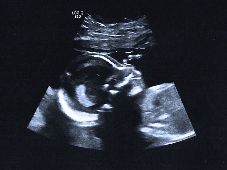
Most medical anatomy scans use 2D ultrasound pictures. These are the classic black-and-white images with bright white outlines, gray shadows, and mysterious blobs that somehow make perfect sense to the sonographer and look like abstract art to everyone else.
Common pictures from the 20-week scan
Profile view: This is often the picture parents recognize most easily. You may see the forehead, nose, lips, and chin in side view. If the baby is in a cooperative mood, this can look surprisingly clear.
Face view: A front-facing image may show the eye sockets, nasal area, and mouth. It can look a little spooky if you are not prepared, because ultrasound sees bones and soft tissue differently from a regular camera. Do not panic if your baby briefly resembles a tiny moon astronaut.
Hands and feet: The scan may capture fingers, toes, or a foot pressed near the uterus. These images are often parent favorites because they feel instantly human and adorable.
Spine view: The spine may appear as a bright curved line or a row of small white segments. This view helps the sonographer evaluate alignment and closure.
Heart view: The heart image may show four chambers moving in real time. It is not always the “cute fridge photo,” but medically, it is one of the most important views.
Gender/sex view: If parents want to know, the sonographer may be able to identify fetal sex based on visible anatomy. However, position matters. Crossed legs, a turned body, or an unhelpful angle can turn the big reveal into a polite “maybe later.”
2D, 3D, and 4D ultrasound: What is the difference?
A standard 20-week ultrasound is usually done in 2D. This provides flat, sectional images that are very useful for medical evaluation. A 3D ultrasound creates still images with more surface detail, often showing facial features more realistically. A 4D ultrasound is essentially 3D imaging shown in motion.
Although 3D and 4D images can be exciting, they are not always part of a routine anatomy scan. Many providers reserve them for specific medical reasons or offer a quick image only if it can be done during the medically indicated exam. Health authorities discourage non-medical “keepsake” ultrasounds that add unnecessary exposure without a clinical purpose. Translation: baby pictures are wonderful, but the uterus is not a photo booth at the mall.
How to prepare for a 20-week ultrasound
Preparation instructions vary by clinic, so follow the directions given by your provider. In many cases, you may be asked to arrive with a partially full bladder, especially if the clinic says it helps improve certain views. Other offices do not require this at 20 weeks. Comfortable two-piece clothing can make the appointment easier because the sonographer usually needs access to the abdomen.
Helpful preparation tips
- Bring your insurance card, ID, and any paperwork requested by the office.
- Ask in advance whether guests are allowed, since office policies vary.
- Decide whether you want to know the fetal sex before the appointment starts.
- Tell the sonographer if you do not want the sex revealed.
- Expect the exam to take longer than a quick heartbeat check.
- Prepare questions, but remember that the sonographer may not be allowed to interpret findings in detail.
What happens during the procedure?
Most 20-week ultrasounds are performed transabdominally. You lie on an exam table, expose your abdomen, and the sonographer applies warm or cool gel to help the transducer glide across the skin. The transducer sends and receives sound waves, creating images on a screen.
The sonographer moves the probe across different areas of your abdomen, presses gently or firmly at times, and captures required images and measurements. You may be asked to shift positions, roll slightly, empty your bladder, or take a short walk if the baby is not in a helpful position. Babies, as it turns out, do not read appointment schedules.
Sometimes, a transvaginal ultrasound may be used to get a better look at the cervix or placenta. This involves placing a slender ultrasound probe into the vagina. It can sound intimidating, but it is commonly used when medically needed and usually provides clearer views of certain structures.
How long does the 20-week ultrasound take?
The scan often takes about 30 to 60 minutes, though it can be shorter or longer. A detailed anatomy scan may take more time if the baby’s position makes certain views hard to capture, if there are twins or multiples, or if the provider needs additional images.
An appointment that runs long does not automatically mean something is wrong. Sometimes the baby is facing the wrong way, curled up, moving constantly, or treating the placenta like a privacy curtain. The sonographer’s job is to collect specific views, and patience is part of the process.
Can the 20-week ultrasound reveal the baby’s sex?
Often, yes. Around 20 weeks, fetal anatomy may be developed enough for the sonographer to identify sex if the baby is positioned well. However, ultrasound sex prediction is not guaranteed. The view may be blocked by fetal position, legs, cord placement, or shadows.
If you want a surprise, say so before the scan begins. Some parents ask the sonographer to write the result down and seal it for a later reveal. Others want to know immediately, because patience is not always included in the pregnancy symptom package.
What if the ultrasound finds something unusual?
If the scan finds something that needs closer review, your provider may recommend a follow-up ultrasound, referral to maternal-fetal medicine, fetal echocardiography, genetic counseling, blood testing, amniocentesis, or another type of evaluation. The next step depends on the specific finding, your pregnancy history, and your provider’s assessment.
It is important to know that an ultrasound finding is not always a diagnosis. Some findings resolve, some are minor, and some need monitoring. Others may help the care team plan for delivery at a hospital with specialized newborn care. The emotional part can be hard, so ask your provider to explain what was seen, how certain they are, what it might mean, and what happens next.
What if the scan is incomplete?
An incomplete anatomy scan means the sonographer could not get all required images. This is common and does not automatically signal a problem. Fetal position, movement, body habitus, abdominal scars, low fluid, or early gestational age can make images harder to capture.
If this happens, your provider may schedule another scan in a week or two. By then, the baby may be in a better position, and structures may be easier to see. In other words, an incomplete scan may simply mean your baby declined to participate in the full photoshoot.
Is a 20-week ultrasound safe?
Medical ultrasound has a strong safety record when performed for a medical reason by trained professionals. It uses sound waves rather than ionizing radiation. Still, major medical organizations recommend using ultrasound prudently: only when medically indicated, for the appropriate amount of time, and by qualified healthcare providers.
That is why many experts advise against non-medical boutique ultrasounds done only for keepsake pictures or videos. The concern is not that a standard anatomy scan is unsafe; the concern is unnecessary exposure, non-clinical use, and the risk that untrained operators may miss or misinterpret findings.
What the 20-week scan cannot do
The anatomy scan is powerful, but it has limits. It cannot detect every birth defect, genetic condition, developmental issue, or future pregnancy complication. Some conditions are too subtle to see on ultrasound, some appear later in pregnancy, and some require other forms of testing.
The scan also does not measure personality, future sleep habits, or whether your child will one day hide a banana in the couch cushions. Science has boundaries.
Questions to ask at your appointment
- Did the scan get all the views needed?
- Is fetal growth measuring as expected?
- Where is the placenta located?
- Is the amniotic fluid level normal?
- Was the heart seen clearly?
- Do I need a follow-up ultrasound?
- Are there any findings I should discuss with my provider?
- Can I receive printed or digital ultrasound pictures?
Real-life experiences: What parents often feel before, during, and after the 20-week ultrasound
The 20-week ultrasound is often described as exciting, but “exciting” is a small word trying to carry a very large emotional backpack. Many parents walk into the appointment feeling thrilled, nervous, curious, and slightly overwhelmed all at once. One minute, they are wondering whether the baby will look like them. The next minute, they are silently hoping every organ, bone, and tiny measurement looks exactly as it should.
A common experience is surprise at how medical the appointment feels. Movies sometimes make ultrasounds look like a five-minute bonding session with instant smiles and perfect pictures. In real life, the room may be dim, the sonographer may be focused, and the screen may show images that are not instantly understandable. Parents may hear quiet clicking, see measurements appear, and wonder whether silence means concern. Usually, it means the sonographer is concentrating. They are collecting images, labeling structures, and making sure the provider has what they need.
Another common experience is laughter. Babies move, stretch, roll, cover their faces, cross their legs, or curl into positions that make the scan more challenging. A parent may arrive hoping for a sweet profile picture and leave with an image that looks like a tiny foot, a mysterious elbow, and possibly a weather map. That is normal. Ultrasound pictures are meaningful, but they are not always refrigerator-ready masterpieces.
Some parents feel emotional when they see the heart beating or the spine in bright white lines. Others feel detached at first because the image is hard to interpret. Both reactions are normal. There is no “correct” way to feel during an anatomy scan. Pregnancy can be tender, strange, funny, and stressful in the same hour.
For parents who have had previous losses, fertility treatment, high-risk pregnancies, or concerning screening results, the 20-week ultrasound can feel especially intense. The appointment may bring hope, but also fear. In that situation, it can help to bring a supportive person if allowed, write down questions in advance, and ask the provider to explain results in plain language. “Everything looks good so far” and “We need one more view” can feel very different, so clarity matters.
Many parents also learn that follow-up scans are more common than they expected. A repeat ultrasound may be recommended because the baby’s position blocked the heart, spine, face, or hands. Hearing “we need another scan” can cause instant worry, but it often means the team simply needs better pictures. The baby may have been facing the wrong direction, tucked low, or having a dramatic main-character moment.
After the scan, parents often replay the appointment in their heads. They look at the printed pictures, text family members, or sit quietly with the experience. If results are normal, there may be relief. If results are uncertain, there may be anxiety while waiting for the next step. The best practical advice is to avoid interpreting ultrasound images alone. Online image searches can turn a normal shadow into a midnight panic spiral. Your care team is the right source for interpretation.
Ultimately, the 20-week ultrasound is both a medical exam and a milestone. It can offer reassurance, provide helpful information, and make the pregnancy feel more real. It may also remind parents that babies are already wonderfully unpredictable. Even before birth, they have opinions about camera angles.
Conclusion
The 20-week ultrasound is one of the most detailed and meaningful prenatal appointments. It gives healthcare providers a close look at fetal anatomy, growth, placenta location, amniotic fluid, and other important pregnancy details. For parents, it can also be the first time the baby looks less like a tiny blur and more like a person with a profile, feet, hands, and possibly a talent for hiding from the camera.
While the scan can identify many concerns, it cannot detect everything. An incomplete scan or follow-up request is not automatically bad news. The best approach is to ask questions, follow your provider’s guidance, and remember that the anatomy scan is one part of a larger prenatal care plan.