Endometriosis can be difficult to diagnose because its symptoms often overlap with other conditions, including ovarian cysts, irritable bowel syndromenstrual cramps. A pelvic MRI can help untangle that diagnostic knot by creating detailed images of the uterus, ovaries, bowel, bladder, ureters, and supporting pelvic tissueswithout using ionizing radiation.
However, MRI is not a magical “yes or no” machine. It is especially useful for identifying ovarian endometriomas and mapping deep endometriosis, but tiny superficial implants may remain invisible. In other words, a normal MRI is reassuring in some ways, yet it does not automatically erase symptoms or rule out every form of the disease.
What Is Endometriosis?
Endometriosis is a chronic condition in which tissue similar to the lining of the uterus grows outside the uterine cavity. Lesions may develop on the ovaries, fallopian tubes, pelvic lining, uterosacral ligaments, bowel, bladder, ureters, diaphragm, or surgical scars. These growths can trigger inflammation, scarring, adhesions, cyst formation, pain, and changes in pelvic anatomy.
Common symptoms include severe menstrual cramps, chronic pelvic pain, pain during or after sex, painful bowel movements, painful urination, bloating, heavy bleeding, fatigue, and infertility. Symptom severity does not always match the visible extent of disease. A person with a small lesion may have intense pain, while someone with extensive endometriosis may have surprisingly mild symptoms.
Modern clinical guidance increasingly supports making a presumptive diagnosis from symptoms, examination, and imaging rather than requiring immediate diagnostic surgery for everyone. Imaging can therefore help patients begin treatment sooner and can identify disease that needs specialized surgical planning. n>
What Is a Pelvic MRI for Endometriosis?
Magnetic resonance imaging uses a powerful magnetic field, radio waves, and computer processing to create high-resolution pictures of soft tissues. Unlike an X-ray or CT scan, MRI does not expose the patient to ionizing radiation.
A routine pelvic MRI and a dedicated endometriosis MRI are not always identical. A dedicated protocol is designed to search systematically for blood products, fibrosis, adhesions, organ distortion, ovarian cysts, and nodules in locations commonly affected by endometriosis. The radiologist may evaluate the pelvis compartment by compartment instead of simply looking for a mass and moving on.
The quality of the exam depends on both the imaging technique and the reader’s experience. Deep bowel or ligament disease can be subtle, and an MRI performed for a different reason may not be optimized to detect it. A dedicated protocol interpreted by a radiologist familiar with endometriosis is often more informative than a generic scan.
When Do Doctors Recommend Pelvic MRI?
Transvaginal ultrasound is commonly used as the first imaging test because it is widely available, relatively inexpensive, and good at detecting ovarian endometriomas. MRI may be ordered when ultrasound findings are unclear, when symptoms and ultrasound results do not match, or when a broader map of the pelvis is needed.
Common reasons for ordering MRI include:
- Suspected deep infiltrating endometriosis involving the bowel, bladder, ureters, vagina, or pelvic ligaments
- Possible ovarian endometriomas or complex adnexal masses
- Persistent symptoms despite a negative or indeterminate ultrasound
- Preoperative mapping before endometriosis surgery
- Evaluation of recurrent pain after previous surgery
- Concern about adenomyosis, fibroids, congenital abnormalities, or another condition that may mimic endometriosis
The American College of Radiology considers pelvic MRI, with or without intravenous contrast depending on the clinical situation, usually appropriate for suspected endometriosis and for further evaluation after an inconclusive ultrasound. MRI is also useful when rectosigmoid involvement is suspected. n>
What Can Pelvic MRI Detect?
Ovarian endometriomas
Endometriomas are cysts filled with old blood, sometimes nicknamed “chocolate cysts”a name that sounds like dessert and absolutely is not. MRI can often distinguish these cysts from other ovarian masses by analyzing their signal patterns on different sequences.
Deep infiltrating endometriosis
Deep endometriosis extends beneath the surface of the pelvic lining and may form fibrotic plaques or nodules. MRI can help locate lesions involving the uterosacral ligaments, rectovaginal space, posterior vaginal fornix, bladder wall, bowel, and other pelvic structures.
Adhesions and distorted anatomy
Endometriosis can cause organs to stick together. MRI may show indirect signs such as ovaries pulled toward the midline, an ovary fixed behind the uterus, a sharply bent rectum, or loss of the normal tissue planes between organs. These clues can be important even when an individual implant is not obvious.
Bowel, bladder, and ureter involvement
When endometriosis affects the bowel, MRI may estimate the lesion’s location, length, depth, and relationship to the bowel wall. It can also identify bladder nodules or ureteral obstruction, which may require collaboration among gynecologic surgeons, colorectal surgeons, or urologists.
Adenomyosis and other pelvic conditions
Adenomyosis occurs when endometrial-type tissue grows within the muscular wall of the uterus. It can coexist with endometriosis and cause heavy bleeding, cramping, and an enlarged or tender uterus. MRI can also reveal fibroids, ovarian cysts, pelvic masses, or structural abnormalities that may explain symptoms.
MRI provides a broad view of the pelvis, while expert ultrasound can offer highly detailed real-time assessment of selected structures. The two tests are often complementary rather than competitors in a diagnostic cage match. n>
What MRI May Miss
The biggest limitation is superficial peritoneal endometriosis. These implants can be tiny, flat, and scattered across the pelvic lining. They may not distort anatomy or contain enough blood or fibrosis to stand out on MRI. As a result, a report that says “no MRI evidence of endometriosis” does not always mean no endometriosis exists.
Accuracy can also vary with lesion location, scanner strength, bowel motion, preparation, protocol design, and radiologist expertise. Hormonal treatment may change the appearance of some lesions, although patients generally should not stop prescribed medication solely for imaging unless their clinician specifically instructs them to do so.
Clinical context remains essential. Persistent cyclical pain, painful bowel movements, deep pain during sex, infertility, or a strong family history may still justify specialist evaluation despite a normal scan. Reviews of endometriosis imaging consistently note that MRI performs best for ovarian and deep disease and is less reliable for superficial implants. n>
How to Prepare for a Pelvic MRI
Preparation varies by imaging center, so follow the facility’s instructions rather than a random checklist from the internet. Depending on the protocol, you may be asked to avoid food for four to six hours, arrive with a moderately full bladder, use a mild bowel preparation, or receive medication that temporarily reduces bowel movement.
Some centers place ultrasound gel in the vagina or rectum to outline pelvic structures more clearly. This is not used everywhere, and patients should be told in advance. You may discuss alternatives if the step is likely to cause distress, pain, or significant discomfort.
Tell the MRI team before the scan if you:
- May be pregnant or are breastfeeding
- Have kidney disease or receive dialysis
- Have a pacemaker, neurostimulator, cochlear implant, aneurysm clip, metal fragment, or other implanted device
- Have previously reacted to MRI contrast
- Have severe claustrophobia, chronic pain, or difficulty lying flat
- Use a medication patch that may contain metal
Remove jewelry, piercings, watches, hearing aids, removable dental devices, and clothing with metal fasteners as instructed. Bring prior ultrasound, CT, MRI, operative, and pathology reports when possible. Older records can help the radiologist understand what has changed and where disease was previously found.
What Happens During the Scan?
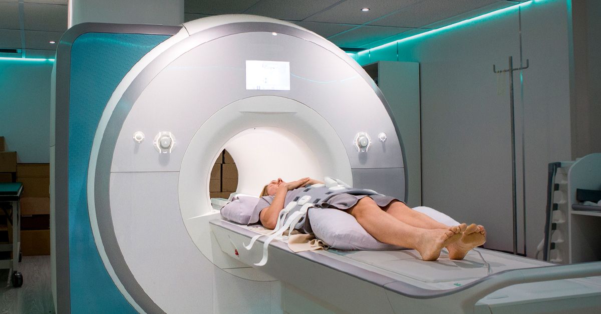
You will usually change into a gown and lie on your back on a padded table. A special receiver called a pelvic coil is positioned over the lower abdomen. The table then moves into the MRI scanner, which is shaped like a large tube.
The technologist communicates through an intercom and can see you throughout the exam. The machine makes loud knocking, thumping, and buzzing sounds, so you will receive ear protection. You must remain as still as possible while each series of images is collected. Some sequences require a brief breath-hold.
A pelvic MRI often lasts about 30 to 60 minutes, although a dedicated endometriosis protocol may take longer. Patients with pain can ask for cushions under the knees, a blanket, or a pause between sequences. The scanner is noisy, not judgmental; it does not care whether your socks match.
MedlinePlus notes that pelvic MRI commonly takes 30 to 60 minutes and does not use ionizing radiation. n>
Will You Need Contrast?
Not every endometriosis MRI requires gadolinium-based contrast. Many characteristic findings can be seen on noncontrast sequences, including T1-weighted images that highlight blood products and T2-weighted images that show anatomy and fibrosis.
Contrast may be helpful when the radiologist needs to characterize an indeterminate mass, assess inflammation, evaluate a suspicious solid component, or distinguish endometriosis from another condition. The decision depends on the imaging protocol and the clinical question.
Allergic reactions to gadolinium are uncommon, but patients with severe kidney impairment require special assessment because certain contrast agents can pose additional risk. Pregnancy should also be discussed before contrast is given. Never assume that “dye” is routine; ask why it is recommended and whether the result would change treatment.
Understanding the MRI Report
Reports may contain unfamiliar anatomical terms. The “anterior compartment” generally includes the bladder and structures in front of the uterus. The “posterior compartment” includes the area behind the uterus, such as the rectum, rectovaginal space, and uterosacral ligaments.
Terms you may see include:
- Endometrioma: An ovarian cyst with imaging features typical of endometriosis
- Deep endometriosis: A nodule or plaque extending beneath the peritoneal surface
- Fibrotic thickening: Scar-like tissue that may reflect chronic disease
- Tethering: Abnormal pulling or fixation of an organ
- Adhesions: Scar tissue connecting structures that normally move separately
- Obliterated cul-de-sac: Scarring that closes the space behind the uterus
- Hydroureter or hydronephrosis: Swelling caused by impaired urine drainage, which may require prompt attention
- No definite MRI evidence: No visible lesion was identified; this is not always the same as complete exclusion
The most useful report should describe lesion location, size, organ involvement, and complications. Ask your clinician how the findings connect to your symptoms and whether the images were interpreted using a dedicated endometriosis approach.
What Happens After the MRI?
A radiologist reviews the images and sends a report to the ordering clinician. Results may appear in a patient portal before your appointment, occasionally introducing readers to phrases that sound like rejected science-fiction villains. Try not to interpret one isolated term without the full clinical context.
Possible next steps include medication for pain or hormonal suppression, pelvic floor physical therapy, fertility evaluation, referral to an endometriosis specialist, additional ultrasound, or surgical consultation. MRI does not determine treatment by itself. Decisions should consider symptom severity, fertility goals, age, prior treatment, lesion location, and patient preferences.
When surgery is being considered, MRI can help the team anticipate whether bowel, bladder, or urinary tract specialists may be needed. That planning can reduce surprises in the operating room and help patients understand the potential scope of the procedure. Mayo Clinic notes that MRI can provide detailed information about the size and location of endometriosis growths for surgical planning. n>
Practical Experiences: What a Pelvic MRI for Endometriosis May Feel Like
The following is a realistic composite of common patient experiences rather than the story of one identifiable person.
The appointment often begins with paperwork that asks the same questions in several creative formats: Do you have metal in your body? Are you sure? Is there any chance an old workplace incident left a metal fragment near your eye? These questions may feel repetitive, but MRI safety screening matters because the scanner uses a powerful magnet.
After changing into a gown, the technologist explains the protocol. Some patients are surprised that preparation differs from what a friend experienced. One person may be asked to fast, another may need a moderately full bladder, and another may receive an antispasmodic medication. Dedicated endometriosis imaging is not completely standardized across facilities, so asking for instructions ahead of time prevents last-minute confusion.
Positioning can be the hardest part for people with pelvic or lower-back pain. Lying flat and still for 45 minutes may sound easy until the pelvis begins protesting like it has filed a formal complaint. A cushion under the knees can reduce strain. Patients should mention pain before the scan begins rather than attempting heroic silence. Technologists can often adjust padding, provide a blanket, or allow a short break between image sets.
The scanner is loud. Earplugs or headphones soften the knocking, but the sound remains noticeable. Many patients cope by counting sequences, focusing on slow breathing, imagining a familiar place, or keeping their eyes closed before the table moves. Those with claustrophobia may benefit from discussing anti-anxiety medication or a wider-bore scanner in advance. If sedation is used, transportation home may be required.
When contrast is ordered, an IV is usually placed in the hand or arm. Some people notice a brief cool sensation during injection, while many feel nothing. Questions about kidney function, pregnancy, prior reactions, and the purpose of contrast are appropriate. Good care includes explanation, not a mysterious syringe appearing without context.
The emotional part can be more difficult than the physical procedure. Patients who have spent years being told that severe period pain is “normal” may hope the MRI finally provides visible proof. A positive scan can feel validating, even though it brings unwelcome news. A normal scan can feel crushing, especially when symptoms are severe. It helps to know beforehand that superficial endometriosis may not appear on MRI. Negative imaging should not become permission to dismiss persistent symptoms.
Afterward, most people can return to normal activities unless they received sedation. The waiting period for results can produce an impressive amount of patient-portal refreshing. When the report arrives, phrases such as “tethering,” “T1 hyperintense focus,” or “posterior compartment disease” may sound alarming. Writing down questions before the follow-up visit can make the discussion more productive.
Useful questions include: Was this a dedicated endometriosis protocol? Were the bowel, bladder, ureters, and uterosacral ligaments specifically assessed? Does the report explain my symptoms? Could superficial disease still be present? Would treatment change based on these findings? Should the images be reviewed by an endometriosis-focused radiologist or surgeon?
The most constructive experience is not necessarily the one with the clearest scan. It is the one in which the patient leaves with a planwhether that means medication, physical therapy, fertility support, specialist review, surgery, or continued investigation. MRI is a powerful map, but it is still one page in the larger medical story.
Conclusion
Pelvic MRI for endometriosis is a valuable, noninvasive tool for detecting endometriomas, identifying deep disease, assessing organ involvement, and planning treatment. It offers excellent soft-tissue detail and a broad view of the pelvis without ionizing radiation. Its greatest strength is mapping disease that may affect surgery or require a multidisciplinary team.
Its greatest limitation is equally important: small superficial lesions may not be visible. A normal MRI should be interpreted alongside symptoms, examination findings, prior imaging, and treatment response. The goal is not merely to obtain a beautiful set of images. The goal is to turn those imagespositive, negative, or uncertaininto a thoughtful next step.